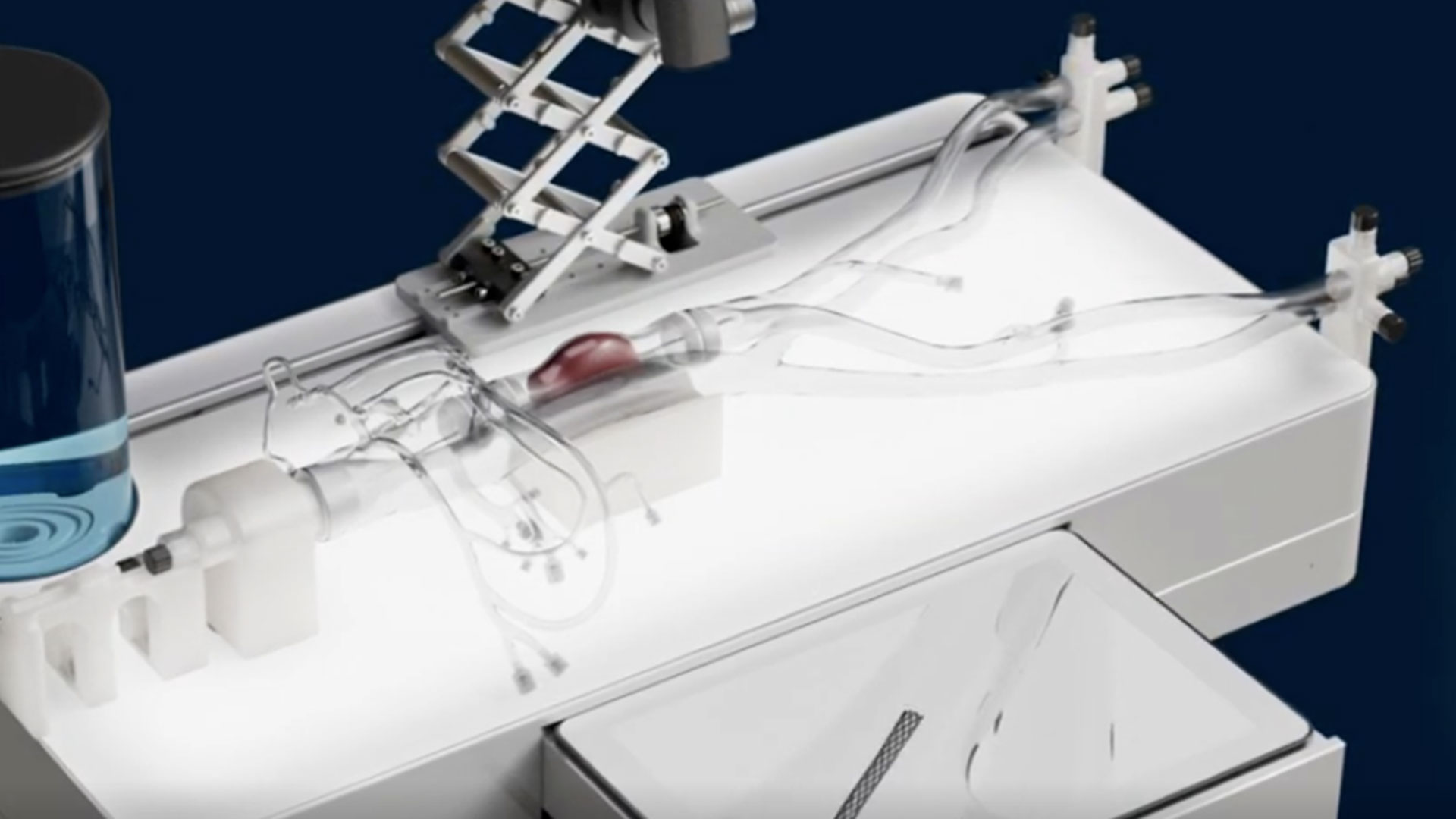
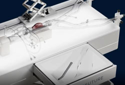
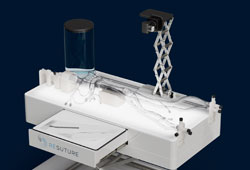
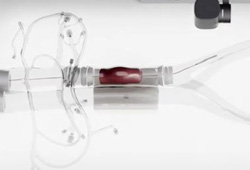
Display screen to track navigation and visualize device performance
Camera system that records every case — the foundation for objective performance metrics
Replaceable components allow for simulation of aneurysm, thrombus, & atherectomy
What is the NAVIS endovascular simulator?
NAVIS — the Navigation and Angio Visualization Integrated Simulator — is an innovative, affordable, and realistic platform built to transform how vascular training is performed today. It is a portable, versatile simulator designed to replicate real-world endovascular procedures using synthetic vasculature and integrated digital visualization.
Where traditional endovascular training depends on cadaver labs, animal labs, or scarce fluoroscopy suite time, NAVIS Lab brings realistic procedural practice to the classroom, the skills lab, or the conference floor. Replaceable, clinically accurate disease states let users practice the full arc of an intervention — catheter and wire navigation, device delivery, and deployment — then reset and repeat as many times as training requires.
Across its disease states, NAVIS Lab supports practice in diagnostic angiography, percutaneous transluminal angioplasty (PTA), stenting, thrombectomy, atherectomy, and aneurysm repair — the core of an endovascular curriculum on a single platform.
Diagnostic angiography
Balloon angioplasty (PTA)
Stenting
Thrombectomy
Atherectomy
Aneurysm repair
Radiation-free fluoroscopy simulation
NAVIS Lab renders live fluoroscopy imagery without a single photon of radiation. A camera captures the trainee’s actual catheter and device movement inside the synthetic vasculature, and the platform’s imaging pipeline converts that feed in real time into the inverted grayscale view an interventionalist expects on the monitor — with anatomical structure blended beneath, so devices are read against recognizable landmarks rather than an empty field.
The workflow is gated the way the real one is. Imaging is driven by a foot pedal: the picture is live only while the trainee is on the pedal, exactly as in a fluoroscopy suite. Trainees build the pedal-and-hands coordination habit — and the instinct to minimize screen time — from the first session, without lead, without a C-arm, and without exposing learners or faculty to any dose at all.
That matters for access as much as for safety. Angio suite time is among the hardest resources for a residency program to schedule and among the most expensive to reserve for training rather than patients. NAVIS Lab decouples endovascular skill development from the fluoroscopy suite entirely.
STEP 1Real wires & devices in synthetic vasculature
STEP 2A camera captures the live movement
STEP 3The pipeline renders a fluoroscopic view
STEP 4A foot pedal gates it — live only on the pedal
Zero radiation · no lead · trains anywhere
Clinically accurate synthetic vasculature
The platform’s replaceable components allow for simulation of aneurysm, thrombus, atherectomy, and stenosis cases, and custom anatomy can be built to match a specific training or device-evaluation need. A single system covers a broad procedural curriculum. Each disease state is built to be clinically accurate rather than merely representative — the anatomy behaves the way the trainee will find it in the patient.
Fluid runs through the synthetic vasculature, so wires and devices track through a wet, flowing field the way they do in a real vessel — not a dry bench model. Because the vessel modules are replaceable, programs are not locked into one scenario: a session can move from a straightforward diagnostic case to a challenging lesion without swapping platforms, and consumable modules can be replaced as they wear rather than retiring the whole simulator.
A visual record of every case — and a path to objective metrics
Because imaging runs off a live camera feed, every run produces a viewable record of how the catheter and device actually moved through the anatomy — reviewable with the trainee instead of reconstructed from memory. Faculty can replay a case, pause on the moment a wire crossed a lesion, and coach against what happened rather than what the trainee remembers.
That visual record is also the foundation for what comes next. ReSuture’s aim is to be the first company capable of delivering quantitative data on surgical performance, and the camera-based platform is being built toward automated performance metrics — objective measures derived from the same footage. That work is grounded in research partnerships with St. Joseph’s Hospital and Medical Center, the Mayo Clinic, and the Arizona State University Department of Machine Learning. Read more about our research and development program.
Who NAVIS Lab is built for
Vascular surgery and interventional residency programs
Give trainees repeatable endovascular practice without competing for angio suite time, and document competency with objective performance data. Explore our full range of surgical training products.
Medical device manufacturers
By replacing cadaver and animal labs, ReSuture helps the medical device industry bring products to the operating room more efficiently and ethically. NAVIS Lab gives device teams a consistent, repeatable environment for physician training, sales demonstrations, and design feedback. See our medical device solutions.
Research partners
Reproducible anatomy and instrumented performance capture make NAVIS Lab a viable research platform for studying technique, learning curves, and device interaction.
$2.6M
Grant-funded research
5
Major research partners
2
Issued patents
100s
Of doctors trained
Research partners
St. Joseph's Hospital & Medical CenterMayo ClinicArizona State UniversityNSF ASSIST CenterArizona WearTech Center
Frequently asked questions
What procedures can be simulated on NAVIS Lab?
NAVIS Lab supports diagnostic angiography, percutaneous transluminal angioplasty (PTA), stenting, thrombectomy, atherectomy, and aneurysm repair. Replaceable disease states — aneurysm, thrombus, atherectomy, and stenosis, plus custom anatomy — let a single platform cover a broad endovascular curriculum.
Does NAVIS Lab use real radiation?
No. NAVIS Lab produces no radiation at all. Fluoroscopic imagery is generated from a live camera feed and rendered in real time to match the appearance of fluoroscopy, so no lead, no shielding, and no dose monitoring are required — for trainees or for faculty.
Does NAVIS Lab use a foot pedal like a real fluoroscopy suite?
Yes. Imaging is gated by a foot pedal, so the image is live only while the pedal is depressed — the same workflow as a real fluoroscopy unit. Trainees build pedal-and-hands coordination, and the habit of minimizing screen time, from the first session.
How is NAVIS Lab different from virtual reality endovascular simulators?
NAVIS Lab pairs physical synthetic anatomy with real fluoroscopic-style imaging. Trainees handle actual wires, catheters, and devices in clinically accurate vasculature and see them rendered on screen as they would in the suite — rather than manipulating instrumented proxies against a purely virtual model. Tactile skill transfers directly.
Is NAVIS Lab portable?
Yes. NAVIS Lab is designed as a portable, versatile simulator that can be set up where training happens — a skills lab, a classroom, or a conference floor — rather than requiring a dedicated suite.
Can NAVIS Lab be used for medical device testing and validation?
Yes. ReSuture’s synthetic models are already used by device manufacturers as an alternative to cadaver and animal labs, and NAVIS Lab extends that to endovascular device evaluation and physician training. Custom anatomy can be built to match a specific device or vessel target.
When will NAVIS Lab be available?
NAVIS Lab is coming soon. To be among the first to get it, join the early adopter list — fill in the form on this page and check the early adopter box, and we’ll be in touch as availability opens up.
Request the NAVIS Lab brochure
Fill in your information to receive a brochure with more detail about NAVIS Lab, or check the box below to join the early adopter program.